
|
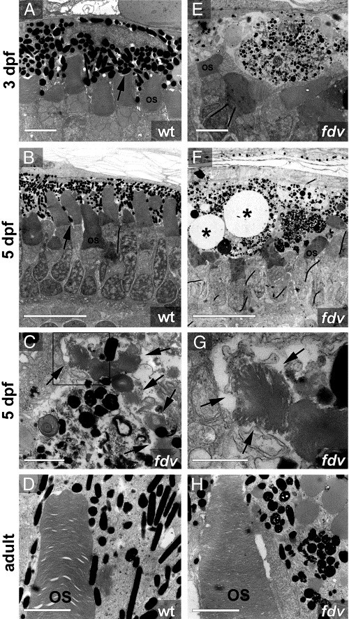
Fig. 3 Transmission electron microscopy from wild-type and fdv. Ultrathin sections were taken from (A, E) 3 dpf, (B, C, F, G) 5 dpf and (D, H) adult zebrafish. At larval stages (A–G), PRC outer segments in fdv mutants are significantly shorter and the RPE is damaged. In the RPE of fdv mutants, vacuoles begin to form at (E) 3 dpf and are prominent at (asterisks in panel F) 5 dpf. Melanin-containing pigment granules are aberrantly shaped. (C and G) Organelles containing detached PRC outer segments can be observed more often within RPE cells of fdv mutants than in wild-type larvae. Panel (G) shows a higher magnification of the box depicted in panel (C). The arrows in panels (C) and (G) indicate the undigested stacks of PRC outer segments in the RPE. The microvilli that interdigitate between the PRC outer segments (arrow in panel B) in wild-type larvae are rarely found in larvae of fdv mutants. (D, H) In adult fdv, the outer segments have recovered, but the melanosomes contain vesicles and some have a fuzzy diffuse shape. OS, outer segments of photoreceptor cells. The thin black lines in panels (B), (E) and (F) are due to preparation artifacts. Scale bars in panels (A) and (E) are 2 μm, in panels (B) and (F) 10 μm, in panel (C) 1 μm, in panel (G) 0,5 μm, in panels (D) and (H) 2 μm.
Reprinted from Developmental Biology, 284(2), Schonthaler, H.B., Lampert, J.M., von Lintig, J., Schwarz, H., Geisler, R., and Neuhauss, S.C., A mutation in the silver gene leads to defects in melanosome biogenesis and alterations in the visual system in the zebrafish mutant fading vision, 421-436, Copyright (2005) with permission from Elsevier. Full text @ Dev. Biol.