
|
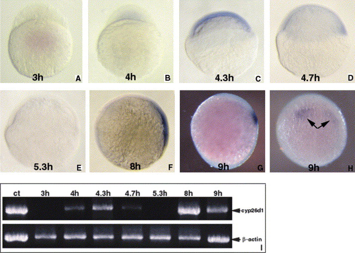
Fig. 2 Analysis of cyp26d1 expression during zebrafish early development from cleavage to gastrulation using in situ hybridization and RT-PCR. (A–E) Animal pole top; (F–G) dorsal right, anterior top; (H) dorsal forward, anterior top. (A) No maternal cyp26d1 message during cleavage is detected in 3 hpf embryos by whole-mount in situ hybridization. (B) Weak expression of cyp26d1 is first found in inner layer of blastula at sphere stage (4 hpf). (C) The expression is found in all blastomeres at dome stage (4.3 hpf). (D–E) Expression of cyp26d1 is decreased at 30% epiboly (4.7 hpf) (D) and cannot be detected at 50% epiboly (5.3 hpf) (E). (F) cyp26d1 resumes its expression in dorsal region at 75% epiboly (8 hf). (G, H) Expression of cyp26d1 is restricted to two separate regions that would presumptively develop into hindbrain at 90% epiboly (9 hpf). Arrows point to the expression areas of cyp26d1 gene. (I) RT-PCR analysis shows the dynamic change of cyp26d1 expression from cleavage to gastrulation of zebrafish embryos. ct, Positive control lane using cyp26d1 cDNA as template; 3–9 h, lanes using total RNA isolated from 3 to 9 hpf as templates; PCR gives a 837 bp amplified fragment from cyp26d1 cDNA. Amplification of a 716 bp β-actin cDNA fragment is a control of RT-PCR sensitivity in the assay. The dynamic changes of cyp26d1 expression from cleavage to gastrulation as detected by RT-PCR is consistent with that detected by whole-mount in situ hybridization.
Reprinted from Gene expression patterns : GEP, 5(6), Gu, X., Xu, F., Wang, X., Gao, X., and Zhao, Q., Molecular cloning and expression of a novel CYP26 gene (cyp26d1) during zebrafish early development, 733-739, Copyright (2005) with permission from Elsevier. Full text @ Gene Expr. Patterns