
|
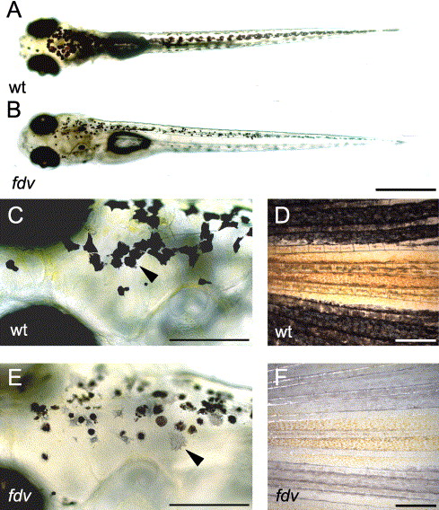
Fig. 1 Body pigmentation of the fading vision mutant. (A) 5-day-old wild-type and (B) fdv mutant larva. fdv shows lighter body pigmentation compared to the 5-dpf-old wild-type larva. (C, E) Higher magnification reveals that the shape of the pigment cells and the distribution of melanin in the pigment cells are different between (C) 5-dpf-old wild-type larvae and (E) 5-dpf-old fdv mutant larvae. (Arrowhead in panels C and E) Some pigment cells appear almost unpigmented. Hypopigmentation is clearly apparent in panel (D) fins of adult wild-type fish compared to (F) homozygous adult fdv mutants. Scale bars in panels (A) and (B) are 500 μm. Scale bars in panels (C) and (E) are 200 μm. Scale bars in panels (D) and (F) are 500 μm.
Reprinted from Developmental Biology, 284(2), Schonthaler, H.B., Lampert, J.M., von Lintig, J., Schwarz, H., Geisler, R., and Neuhauss, S.C., A mutation in the silver gene leads to defects in melanosome biogenesis and alterations in the visual system in the zebrafish mutant fading vision, 421-436, Copyright (2005) with permission from Elsevier. Full text @ Dev. Biol.