
|
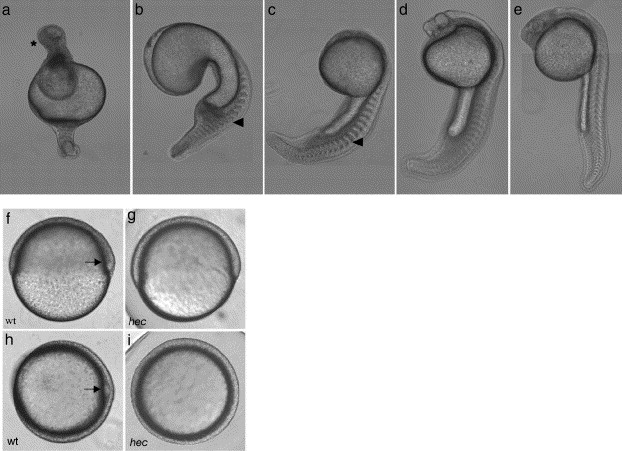
Fig. 1 hecate mutant embryos display defects in axis induction at 24 hpf. (a–e) At 24 hpf, the majority of embryos from a given clutch have severe radial ventralization (a) while the remainder exhibit varying degrees of deficiency in dorsoanterior development (b–e). Anterior: top; posterior: bottom; dorsal: right, when identifiable. In panel a, the anterior contains a mass of apparently undifferentiated cells (asterisk). Blocky somites (arrowheads in panels b and c) are characteristic of the absence of a notochord. (f–i) Wild-type (f, h) and hec mutant (g, i) embryos at the shield stage (6 hpf). Wild-type embryos display a characteristic thickening on the dorsal side (arrows in panels f and h) while hec mutant embryos lack this thickening. (f and g: side views; h and i: animal views).
Reprinted from Developmental Biology, 286(2), Lyman Gingerich, J., Westfall, T.A., Slusarski, D.C., and Pelegri, F., hecate, a zebrafish maternal effect gene, affects dorsal organizer induction and intracellular calcium transient frequency, 427-439, Copyright (2005) with permission from Elsevier. Full text @ Dev. Biol.