
|
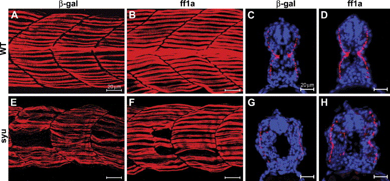
Fig. 7 Partial restoration of slow muscle myofibril organization in syu mutants by transient expression of ff1a. Slow myofibrils were detected by antibody F59. (A, B, E, F) Lateral view of somites 14–17, (C, D, G, H) cross-sections. The blue color is Hoechst dye staining for nuclei. (A) Normal slow muscle fibrils in the β-gal-injected wild-type (WT) embryo at 24 hpf. (B) Overexpression ff1a led to thicker slow myofibrils. (E) Distorted appearance of slow myofibrils in the syu mutant. (F) In the ff1a overexpressed syu mutant, the slow myofibrils are better organized and the width of the myofibril expanded. Scale bars are 20 μm.
Reprinted from Developmental Biology, 286(2), Sheela, S.G., Lee, W.C., Lin, W.W., and Chung, B.C., Zebrafish ftz-f1a (nuclear receptor 5a2) functions in skeletal muscle organization, 377-390, Copyright (2005) with permission from Elsevier. Full text @ Dev. Biol.