
|
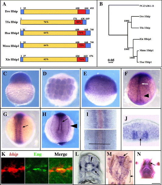
Fig. 1 Adaxial cells, muscle pioneer and slow muscle precursors express hhip mRNA. (A, B) Zebrafish Hhip protein is closely related to Hhip in other vertebrates. (A) The predicted amino acid sequences of Danio rerio (Dre, DQ177323), Homo sapiens (Hsa, NM_022475), Mus musculus (Mmu, AF116865), Xenopus laevis (Xle, BC046952) and Takifugu rubripes (Tfu, SINFRUT00000133748) Hhip protein. Red indicates the EGF-like domains, and blue indicates the hydrophobic stretches. Percentages indicate sequence identity of amino acids of each domain compared to the zebrafish sequence. The numbers indicate the locations of borders between domains. (B) Phylogenetic tree comparing zebrafish Hhip with other vertebrate Hhip proteins. The tree is based on the amino acid sequences of putative open reading frames of the proteins aligned with the Clustal method. PCZA361.11 (CAA11769) was used as an outgroup. Numbers indicate bootstrap support for the nodes. (C) Maternal hhip mRNA is present in the one-cell stage embryo. (D, E) hhip mRNA is present throughout the embryo at 32-cell (D) and 50% epiboly (E) stages. (F) hhip is expressed at higher levels in the midline (arrow) and adaxial cells (arrowhead) at bud stage. (G, H) hhip expression is apparent in the medial somite (G, arrow), adaxial cells (H, arrow) and pronephric tissue (H, arrowhead) at the 8-somite stage. (I) Comparison of hhip (blue) and shh (red) expression at the 12-somite stage. hhip mRNA is expressed adjacent to shh expressing cells in posterior, presomitic regions (bottom) and farther lateral in anterior, segmented regions (top). Bar indicates location of section shown in panel J. (J) hhip is expressed adjacent to the notochord (arrow, notochord). (K) Muscle pioneer cells express hhip mRNA (left panel) and Eng protein (middle panel). Double labeling with 4D9 (green) anti-Eng antibody and hhip (red) shows that hhip expressing cells contain Eng protein (right panel) at 24 hpf. (L) hhip mRNA is detected in a subset of fast muscle cells (arrow) at 24 hpf. (M) hhip is expressed in the tectum (arrow) and neural crest cells (bracket) at 24 hpf. (N) hhip expression in adductor mandibulae at 48 hpf (arrow). (C, E) Lateral views; (D, F, G, M) dorsal views; (H) posterior view of the tail bud; (J, L) transverse sections, dorsal towards the top; (K) lateral view; (N) ventral view. Scale bar: (C–H, K, M, N) 200 μm; (I, J, L) 50 μm; (K) 25 μm.
Reprinted from Developmental Biology, 297(1), Ochi, H., Pearson, B.J., Chuang, P.T., Hammerschmidt, M., and Westerfield, M., Hhip regulates zebrafish muscle development by both sequestering Hedgehog and modulating localization of Smoothened, 127-140, Copyright (2006) with permission from Elsevier. Full text @ Dev. Biol.