
Image
|
Figure Caption
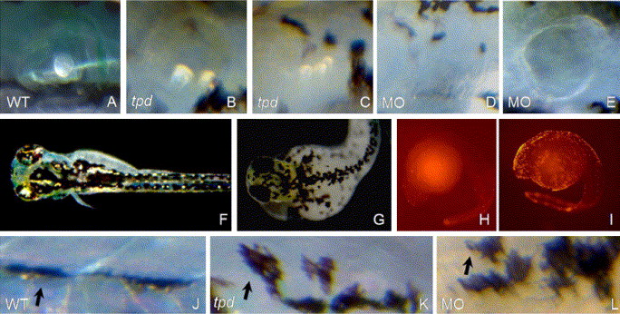
Fig. 3 Ear and pigmentation phenotypes in tpd mutants and Trap230 morphants. (A–G, J–L) Embryos at 4 dpf. (H, I) Embryos at 28 hpf. (A–E) Ear phenotypes; note partial reduction in tpd mutants (C), and complete absence in some Trap230 morphants (D). (F, G) Dark field microscopy, revealing iridophores (bright shiny cells), present in WT (F), but not in tpd mutants (G). (H, I) TUNEL staining, lateral view of embryos. (J–L) Trunk melanocytes (dark cells, arrows). (A, F, H, J) WT; (B, C, G, I, K) tpd mutants; (D, E, L) Trap230 MO-injected embryos.
Acknowledgments
This image is the copyrighted work of the attributed author or publisher, and
ZFIN has permission only to display this image to its users.
Additional permissions should be obtained from the applicable author or publisher of the image.
Reprinted from Developmental Biology, 296(1), Rau, M.J., Fischer, S., and Neumann, C.J., Zebrafish Trap230/Med12 is required as a coactivator for Sox9-dependent neural crest, cartilage and ear development, 83-93, Copyright (2006) with permission from Elsevier. Full text @ Dev. Biol.