
|
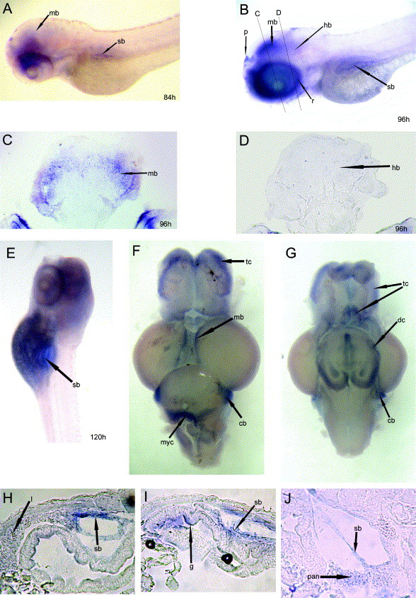
Fig. 3 Expression of DEC1 in early larva stages of zebrafish and in the adult brain. All panels show a lateral view except (F) (dorsal view) and G (ventral view). (C and D) Transverse sections through the midbrain and hindbrain of the 96 hpf embryos, as indicated in (B), respectively. Panels (F–H) are longitudinal sections through the swim bladder of 120 hpf embryos, with the anterior towards the left, and dorsal towards the top. (A) signals in midbrain and swim bladder at 84 hpf. (B–D) DEC1 expression is strong in the midbrain but weak in the hindbrain at 96 hpf. Meanwhile, the strong expression in the pineal gland remained and expression in the eyes was enhanced in the retinal. (E and H–J) The expression in the midbrain declined, but emerged in the swim bladder and digestive system including gut, liver and pancreas at 120 hpf. (F–G) DEC1 expression in the adult brain is mainly restricted in the anterior–lateral telencephalon, the most medial midbrain, and two lateral areas of the cerebellum, ventral diencephalons and myelencephalon. Abbreviations: dc, diencephalons; g, gut; hb, hindbrain; ht, hypothalamus; l, liver; mb, midbrain; myc, myelencephalon; p, pineal gland; pan, pancreas; r, retinal; sb, swim bladder; tc, telencephalon.
Reprinted from Gene expression patterns : GEP, 6(8), Yao, J., Wang, L., Chen, L., Zhang, S., Zhao, Q., Jia, W., and Xue, J., Cloning and developmental expression of the DEC1 ortholog gene in zebrafish, 919-927, Copyright (2006) with permission from Elsevier. Full text @ Gene Expr. Patterns