
|
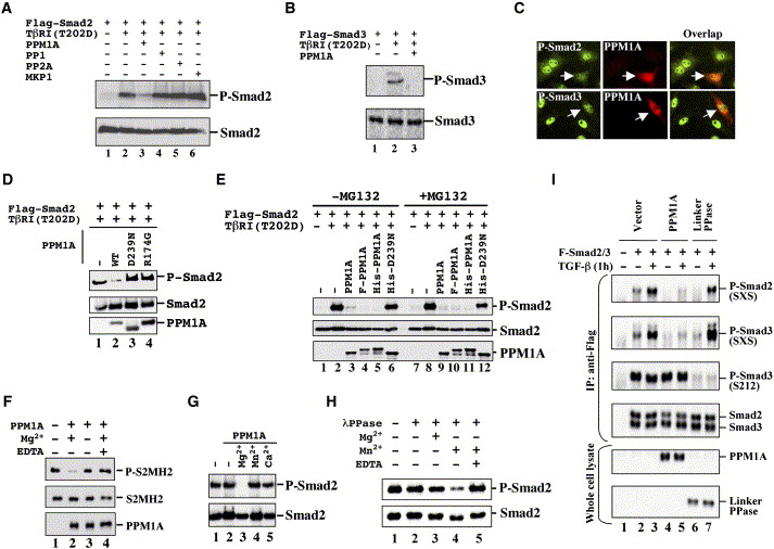
Fig. 2 PPM1A Dephosphorylates Smad2 and Smad3 (A) Smad2 dephosphorylation by PPM1A. In transfected 293T cells, Flag-Smad2 was anti-Flag immunoprecipitated, and levels of P-Smad2 and total Smad2 were assessed by Western blotting. (B) PPM1A dephosphorylates Smad3 in 293T cells. (C) PPM1A reduces endogenous P-Smad2/3 levels. HaCaT cells were transfected with PPM1A, treated with TGFβ (2 ng/ml, 1 hr), and immunostained using anti-PPM1A (Texas red) and P-Smad2 or P-Smad3 (FITC). Arrow indicates reduced P-Smad2/3 in PPM1A-transfected cells. (D) Smad2 dephosphorylation requires the catalytic activity of PPM1A. (E) Smad2 dephosphorylation occurs in the presence of MG-132. Flag-PPM1A or His-PPM1A is as efficient as PPM1A. (F) PPM1A dephosphorylates P-Smad2 MH2 domain (P-S2MH2). Dephosphorylation of P-S2MH2 required 20 mM Mg2+ (lane 2) and was abolished by 40 mM EDTA (lane 4). Equal amounts of semisynthetic recombinant P-S2MH2 (100 ng, lanes 1–4) and recombinant PPM1A (100 ng, lanes 2–4) were loaded. (G) Smad2 dephosphorylation by PPM1A depends on 20 mM Mg2+ (lane 3), but not 2 mM Mn2+ (lane 4) or 20 mM Ca2+ (lane 5). (H) Smad2 dephosphorylation by λ phosphatase (λPPase). (I) PPM1A specifically dephosphorylates the SXS motif, but not pS212 of Smad3. Linker phosphatase specifically dephosphorylates pS212, but not the SXS motif.
Reprinted from Cell, 125(5), Lin, X., Duan, X., Liang, Y.Y., Su, Y., Wrighton, K.H., Long, J., Hu, M., Davis, C.M., Wang, J., Brunicardi, F.C., Shi, Y., Chen, Y.G., Meng, A., and Feng, X.H., PPM1A functions as a Smad phosphatase to terminate TGFbeta signaling, 915-928, Copyright (2006) with permission from Elsevier. Full text @ Cell