
|
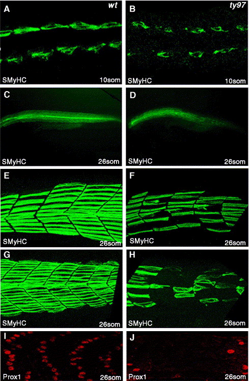
Fig. 2 Slow muscle formation is disrupted in homozygous youmutants. (A–H) Slow myosin heavy chain protein (SMHC) is reduced compared to wild type panels A, C, E, G in youty97 homozygote mutant embryos panels B, D, F, H at 10-somite (A, B) and 26-somite stages (C–H). Panels E, F anterior somites, (G, H) posterior, tail extension somites. (E–H) 3D confocal projections of left sided myotomes. (I, J) At 26 somites, Prox1 protein, a specific marker of slow muscle cells, is reduced or absent compared to wild type (I) within the myotome of youty97 homozygous embryos (J). A–B dorsal confocal sections, at the level of the notochord, anterior to the left. (C–J) Lateral views anterior to the left.
Reprinted from Developmental Biology, 294(1), Hollway, G.E., Maule, J., Gautier, P., Evans, T.M., Keenan, D.G., Lohs, C., Fischer, D., Wicking, C., and Currie, P.D., Scube2 mediates Hedgehog signalling in the zebrafish embryo, 104-118, Copyright (2006) with permission from Elsevier. Full text @ Dev. Biol.