
|
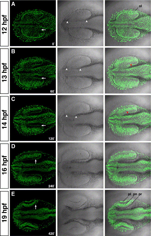
Fig. 8 The prospective RPE is formed by the coordinated migration of the midbrain. (A) At 12 hpf, the optic lobes are fully formed and attached along their A/P axis to the diencephalon (arrowheads in the middle panels). (B) At 13 hpf, the furrow that separates the optic lobes from the diencephalon is formed by the massive migration of the midbrain towards the forebrain region. (C) The furrow is almost fully formed at 14 hpf. The size of the furrow is similar between panels C with D. Therefore, from 14 hpf onwards, the velocity of the midbrain migration decreases drastically. (E) At 19 hpf, the optic cups are formed, and the lens start invaginating from the ectoderm. At all times examined, extraocular mesenchyme is closely associated to the prospective RPE (red arrowheads in panels B and C). The time is given in minutes. Left panels, H2A:H2A-EGFP; middle panels, DIC; right panels, overlay. Anterior is to the left. ol; optic lobe; pl, prospective lens; pn; prospective neural retina; pr, prospective retinal pigment epithelium.
Reprinted from Developmental Biology, 288(2), Rojas-Munoz, A., Dahm, R., and Nüsslein-Volhard, C., chokh/rx3 specifies the retinal pigment epithelium fate independently of eye morphogenesis, 348-362, Copyright (2005) with permission from Elsevier. Full text @ Dev. Biol.