
|
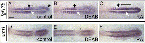
Fig. 9 Expression of genes involved in Fgf-signalling is regulated by RA. 10-somite stage embryos displayed as in Fig. 2. (A–C) In wild-type embryos, fgf17b is expressed in somites, anterior psm (bracket; black arrow indicates position of posterior border of last formed somite), tailbud (arrowhead) and adaxial cells (white arrow; A). In DEAB-treated embryos, fgf17b is reduced in psm, somites and adaxial cells (B); in RA-treated embryos, expression extends to a larger part of anterior psm (bracket) and is increased adaxial cells (white arrow; C). (D–F) In wild-type embryos, erm1 is expressed in tailbud, posterior psm and in somites, but is absent from anterior psm (bracket in panel D); in DEAB-treated embryos, erm1 expression is reduced in somites and posterior psm (E); in RA-treated embryos, erm1 is induced in anterior psm (F). Scale bar: in panel A, 100 μm for panels A–F.
Reprinted from Developmental Biology, 289(1), Hamade, A., Deries, M., Begemann, G., Bally-Cuif, L., Genet, C., Sabatier, F., Bonnieu, A., and Cousin, X., Retinoic acid activates myogenesis in vivo through Fgf8 signalling, 127-140, Copyright (2006) with permission from Elsevier. Full text @ Dev. Biol.