
Image
|
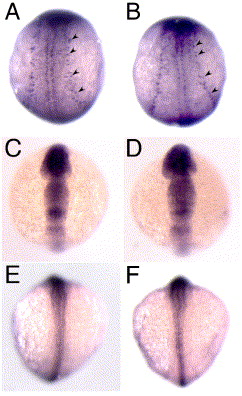
Figure Caption
Fig. S4 Normal development of Rohon–Beard sensory neurons, neural plate and neural plate border in sym1 mutants. Dorsal views of wild-type (A, C, E) and sym1 (B, D, F) mutant embryos. (A, B) huC expression in 3-somite stage embryos show Rohon–Beard sensory neurons (arrowheads) in wild-type and sym1 mutant embryos. (C–F) The ectodermal neural plate and neural plate border domains, are respectively revealed by sox2 expression at the 6-somite stage (C, D) and msxb expression at the 14-somite stage (E, F) in wild-type and sym1 mutant embryos.
Figure Data
Acknowledgments
This image is the copyrighted work of the attributed author or publisher, and
ZFIN has permission only to display this image to its users.
Additional permissions should be obtained from the applicable author or publisher of the image.
Reprinted from Developmental Biology, 292(1), Stewart, R.A., Arduini, B.L., Berghmans, S., George, R.E., Kanki, J.P., Henion, P.D., and Look, A.T., Zebrafish foxd3 is selectively required for neural crest specification, migration and survival, 174-188, Copyright (2006) with permission from Elsevier. Full text @ Dev. Biol.