
Image
|
Figure Caption
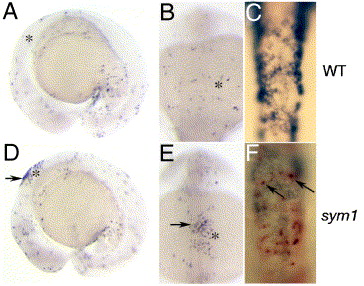
Fig. 8 Neural crest cell death in sym1 mutants. Lateral (A, D) and dorsal (B, C, E, F) views of TUNEL labeled 15-somite stage wild-type (A–C) and sym1 mutant (D–F) embryos shows an increase in dorsal TUNEL-positive cells in the hindbrain region in sym1 mutants (arrows in D, E). Asterisks indicate the position of the developing otic placode. (C, F) Double labeling analysis with ctn (dark blue) and TUNEL (red) shows cell death in some neural crest cells that still express ctn in sym1 mutants at this stage (arrows).
Acknowledgments
This image is the copyrighted work of the attributed author or publisher, and
ZFIN has permission only to display this image to its users.
Additional permissions should be obtained from the applicable author or publisher of the image.
Reprinted from Developmental Biology, 292(1), Stewart, R.A., Arduini, B.L., Berghmans, S., George, R.E., Kanki, J.P., Henion, P.D., and Look, A.T., Zebrafish foxd3 is selectively required for neural crest specification, migration and survival, 174-188, Copyright (2006) with permission from Elsevier. Full text @ Dev. Biol.