
|
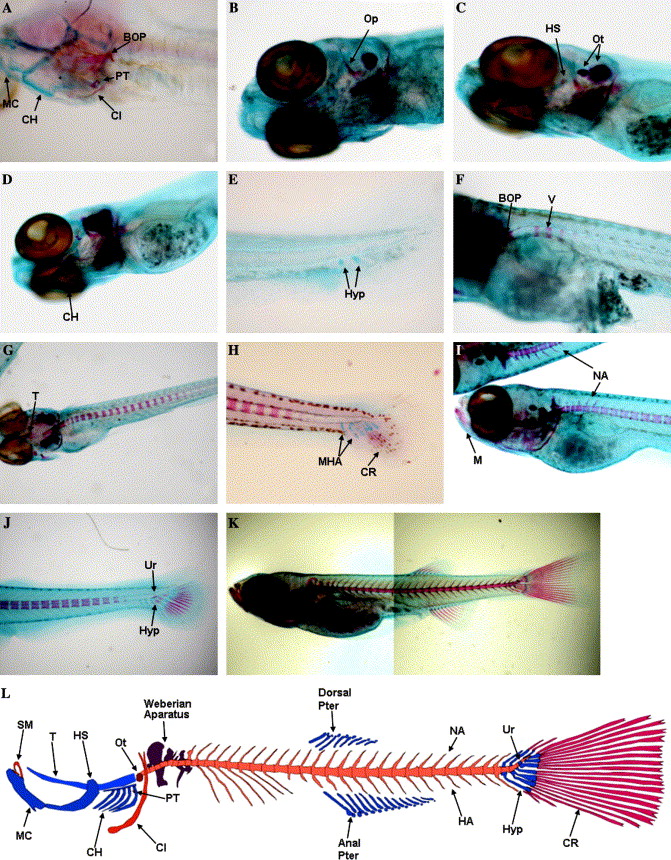
Fig. 1 Time-course of skeletal development in zebrafish using Alcian blue-Alizarin red double staining. Whole mount double staining of the skeleton was used to follow ontogenic development of cartilaginous and calcified structures. (A) 96 hpf zebrafish larvae head skeleton presenting calcified pharyngeal teeth (PT), cleithrum (Cl), and basioccipital articulatory process (BOP) while other structures like Meckel′s cartilage (MC) and ceratohyal (CH) remain as cartilage (100x); (B) 5 dpf zebrafish larva presenting calcification on the opercula (Op) (40x); (C) beginning of the calcification of the hyosymplectic (HS) in a 6 dpf larva (100x); (D) Beginning of the calcification of the ceratohyal (CH) in a 8 dpf zebrafish (100x); (E) First hypurals (Hyp) developing at 8 dpf (100x); (F) The formation of the first vertebrae (V) is observed at 9 dpf (100x); (G) Calcification of the trabeculae (T) is observed at 11 dpf, vertebrae continue to form (in an anterior-posterior direction) towards the posterior end of the notochord (40x); (H) Caudal hypuralia acquires final number of structures with modified hemal arches (MHA) and caudal fin rays (CR) appearing at 12 dpf (100x); (I) Formation of the first neural arches (NA) is observed dorsally in the anterior vertebrae at 14 dpf; note that the mandibular is already calcified (M) (40x); (J) beginning of calcification of the hypurals (Hyp) under the urostile (Ur) at 14 dpf (40x); (K) composite image of a 19 dpf D. rerio with most skeletal structures formed and calcified; (L) schematic representation of the major zebrafish skeletal elements focused on this study. (SM) Supra-mandibular, (PT) pharyngeal teeth, (Ot) otolith, and (HA) hemal arches.
Reprinted from Gene expression patterns : GEP, 6(6), Gavaia, P.J., Simes, D.C., Ortiz-Delgado, J.B., Viegas, C.S., Pinto, J.P., Kelsh, R.N., Sarasquete, M.C., and Cancela, M.L., Osteocalcin and matrix Gla protein in zebrafish (Danio rerio) and Senegal sole (Solea senegalensis): Comparative gene and protein expression during larval development through adulthood, 637-652, Copyright (2006) with permission from Elsevier. Full text @ Gene Expr. Patterns