
Image
|
Figure Caption
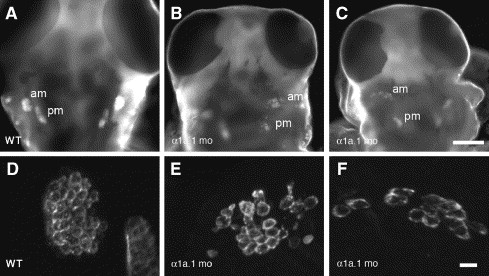
Fig. 8 Macular sensory organs develop in α1a.1 morphants. Hair cells were labeled with HCS-1 antibody in wild type (WT) and α1a.1 morphant embryos at 75 hpf to visualize the anterior (am) and posterior (pm) maculae. (A–C) Low power images of embryos, dorsal views. Scale bar: 100 μm. (D–F) High power views of embryos showing labeled anterior maculae. Scale bar: 10 μm. (A, D) WT embryo. (B, C, E, F) α1a.1 morphants. Both pairs of maculae are present, but they have fewer hair cells and are less tightly clustered than WT. mo, morphant.
Acknowledgments
This image is the copyrighted work of the attributed author or publisher, and
ZFIN has permission only to display this image to its users.
Additional permissions should be obtained from the applicable author or publisher of the image.
Reprinted from Developmental Biology, 294(1), Blasiole, B., Canfield, V.A., Vollrath, M.A., Huss, D., Mohideen, M.A., Dickman, J.D., Cheng, K.C., Fekete, D.M., and Levenson, R., Separate Na,K-ATPase genes are required for otolith formation and semicircular canal development in zebrafish, 148-160, Copyright (2006) with permission from Elsevier. Full text @ Dev. Biol.