
Image
|
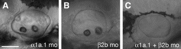
Figure Caption
Fig. 6 Coinjection of Na,K-ATPase α1a.1 and β2b MOs. Embryos were injected with sub-effective doses of α1a.1 and β2b MOs. Lateral views of otic vesicle (OV) at 45 hpf, anterior to the left. (A) OV of embryo injected with 0.125 ng of α1a.1 MO-1. No effect on otolith formation was observed. (B) OV of embryo injected with 2 ng of β2b MO-1 showing normal otolith formation. (C) Embryo co-injected with 0.125 ng of α1a.1 MO-1 and 2 ng of β2b MO-1 failed to form otoliths. mo, morphant. Scale bar: A–C, 50 μm.
Acknowledgments
This image is the copyrighted work of the attributed author or publisher, and
ZFIN has permission only to display this image to its users.
Additional permissions should be obtained from the applicable author or publisher of the image.
Reprinted from Developmental Biology, 294(1), Blasiole, B., Canfield, V.A., Vollrath, M.A., Huss, D., Mohideen, M.A., Dickman, J.D., Cheng, K.C., Fekete, D.M., and Levenson, R., Separate Na,K-ATPase genes are required for otolith formation and semicircular canal development in zebrafish, 148-160, Copyright (2006) with permission from Elsevier. Full text @ Dev. Biol.