
|
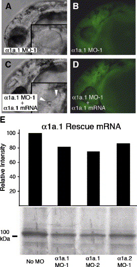
Fig. 2 mRNA rescue of α1a.1 morphant. Coinjection of 125 pg of α1a.1 rescue mRNA and 0.25 ng of α1a.1 MO-1 into one-cell stage embryos. All panels show a lateral view with anterior to the left. (A) α1a.1 morphant at 36 hpf injected with 0.25 ng of α1a.1 MO-1 alone. Inset is an enlarged view of the otic vesicle (OV) lacking otoliths. (B) Fluorescent image of panel A confirming presence of FITC-labeled morpholino in embryos. (C) 36 hpf embryo coinjected with α1a.1 MO-1 (0.25 ng MO-1) and α1a.1 rescue mRNA (125 pg). Inset is an enlarged view of the OV containing two otoliths. Arrowheads indicate otoliths. (D) Fluorescent image of panel C. (E) Effect of MOs on translation of α1a.1 rescue mRNA. The region adjacent to the initiating ATG of α1a.1 rescue mRNA was engineered to contain a minimal Kozak consensus sequence so as not to match the targeting MO. The α1a.1 rescue mRNA was translated in the presence of 4 μM antisense MOs and analyzed as described in Fig. 1A.
Reprinted from Developmental Biology, 294(1), Blasiole, B., Canfield, V.A., Vollrath, M.A., Huss, D., Mohideen, M.A., Dickman, J.D., Cheng, K.C., Fekete, D.M., and Levenson, R., Separate Na,K-ATPase genes are required for otolith formation and semicircular canal development in zebrafish, 148-160, Copyright (2006) with permission from Elsevier. Full text @ Dev. Biol.