
|
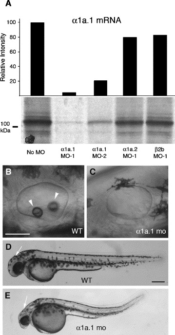
Fig. 1 Na,K-ATPase α1a.1 MOs block otolith formation. (A) Effect of antisense morpholinos on α1a.1 mRNA translation. α1a.1 mRNA was translated in vitro using a rabbit reticulocyte lysate system in the presence of 4 μM antisense MOs. [35S]-methionine-labeled in vitro translation products were analyzed on a 10% SDS-polyacrylamide gel (lower panel). Molecular weight marker is shown at the left. Intensity of the bands relative to control was quantified by densitometry (upper panel). (B-E) Morphants were injected with 0.25 ng of α1a.1 MO-1. Lateral view with anterior to the left. (B) Otic vesicle (OV) of wild type (WT) embryo at 45 hpf. (C) OV of α1a.1 morphant at 45 hpf. (D) WT embryo at 45 hpf. (E) α1a.1 morphant at 45 hpf. Arrowheads indicate otoliths. Arrow indicates midbrain-hindbrain boundary. mo, morphant. Scale bars: A-B, 50 μM; C-D, 250 μM.
Reprinted from Developmental Biology, 294(1), Blasiole, B., Canfield, V.A., Vollrath, M.A., Huss, D., Mohideen, M.A., Dickman, J.D., Cheng, K.C., Fekete, D.M., and Levenson, R., Separate Na,K-ATPase genes are required for otolith formation and semicircular canal development in zebrafish, 148-160, Copyright (2006) with permission from Elsevier. Full text @ Dev. Biol.