
|
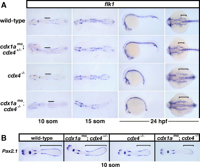
Fig. 3 Expression of flk1 and pax2.1 in cdx-deficient embryos. (A) Whole mount in situ hybridizations showing flk1 transcripts (purple) in cdx-deficient embryos and wild-type controls at the 10- and 15-somite stages (flatmounted embryos shown in dorsal views with anterior to the left) and at 24 hpf (shown in lateral and dorsal views with anterior to the left). Lines mark the rostral angioblast populations. Brackets indicate the regions of flk1-expressing cells near the developing Duct of Cuvier. (B) Whole mount in situ hybridizations showing pax2.1 expression (purple) in the pronephric progenitors (bracket) of wild-type and cdx-deficient embryos at the 10-somite stage. Flatmounted embryos are shown in dorsal views with anterior to the left.
Reprinted from Developmental Biology, 292(2), Davidson, A.J., and Zon, L.I., The caudal-related homeobox genes cdx1a and cdx4 act redundantly to regulate hox gene expression and the formation of putative hematopoietic stem cells during zebrafish embryogenesis, 506-518, Copyright (2006) with permission from Elsevier. Full text @ Dev. Biol.