
|
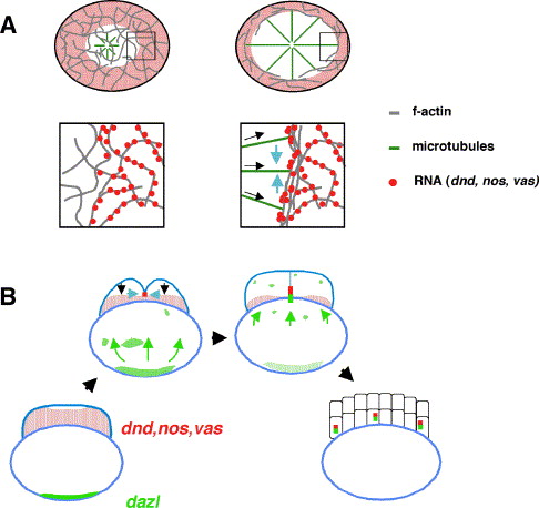
Fig. 8 Models for the recruitment of germ plasm RNAs at the furrow. (A) Cytoskeletal rearrangements and processes involved in the recruitment of early animal germ plasm RNA at the furrow. Diagrams show overview (top) and high magnification (bottom) views of embryos immediately after fertilization (left) and immediately prior to furrow formation (right). Embryos are drawn as animal views. RNA aggregates containing dnd, nos1, and vas RNAs are bound to a network of initially randomly oriented f-actin, forming a prepattern such that the central most region of the blastodisc is RNA-free. As development proceeds, the coordinated growth of astral microtubules (black arrows in bottom right inset) leads to the movement towards the periphery and circumferential alignment of f-actin. Aligned f-actin facilitates aggregation of RNA-containing particles. In a process whose mechanistic basis has not yet been addressed, lateral movement of aggregates in regions immediately adjacent to the forming furrow (blue arrows in bottom right inset) leads to the recruitment of aggregates at the furrow (not shown). (B) Relation of separate pathways of RNA segregation and the compartmentalization of the germ plasm. Embryos are drawn as side views, representing, from bottom left clockwise, a freshly laid egg, an embryo at an early stage of cellularization, an embryo at a late stage of cellularization, and an early blastula embryo. Early animal RNAs such as dnd, nos1, and vas (represented by red dots in stippled region), present in the animal cortex of the freshly laid egg undergo aggregation by the cytoskeletal rearrangements shown in (A) (black arrows) and recruitment at the furrow (large red aggregates) by proposed local lateral transport (blue arrows—see (A)). Vegetally localized RNAs such as dazl and presumably bruno-like reach the distal end of the forming aggregate at the furrow by translocation through the cortex (green arrows). These two mechanisms lead to a compartmentalized structure of the germ plasm, which is maintained during the early cleavage stages.
Reprinted from Developmental Biology, 292(1), Theusch, E.V., Brown, K.J., and Pelegri, F., Separate pathways of RNA recruitment lead to the compartmentalization of the zebrafish germ plasm, 129-141, Copyright (2006) with permission from Elsevier. Full text @ Dev. Biol.