
Fig. 4
- ID
- ZDB-IMAGE-070914-14
- Publication
- Theusch et al., 2006 - Separate pathways of RNA recruitment lead to the compartmentalization of the zebrafish germ plasm
- All Figures
- Figures for Theusch et al., 2006
|
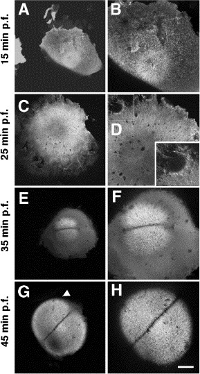
Fig. 4 Microtubule asters emanate from the center of the embryo during the early cellular cycles. Animal views of embryos fixed at progressively older stages of development (A, B: 15 min p.f.; C, D: 25 min. p.f.; E, F: 35 min p.f.; G, H: 45 min p.f.) and labeled with an anti-α-tubulin antibody. Left column: overview; right column, higher magnification views. The insert in panel D shows a higher magnification of the region labeled with a square. (A, B) Immediately after fertilization, a monoastral structure forms at the animal pole of the blastodisc. (C, D) Microtubules of this monoaster increase in length and reach the periphery of the blastodisc, which also exhibits local accumulations of shorter microtubules (inset). (E, F) Immediately prior to the first cleavage, a diastral structure flanking the prospective furrow. (G, H) As in the previous cycle, microtubules of this diaster increase to occupy more peripheral regions of the blastodisc. Scale bar in panel (H) represents 200 μm (left column) and 100 μm (right column).
Reprinted from Developmental Biology, 292(1), Theusch, E.V., Brown, K.J., and Pelegri, F., Separate pathways of RNA recruitment lead to the compartmentalization of the zebrafish germ plasm, 129-141, Copyright (2006) with permission from Elsevier. Full text @ Dev. Biol.