
|
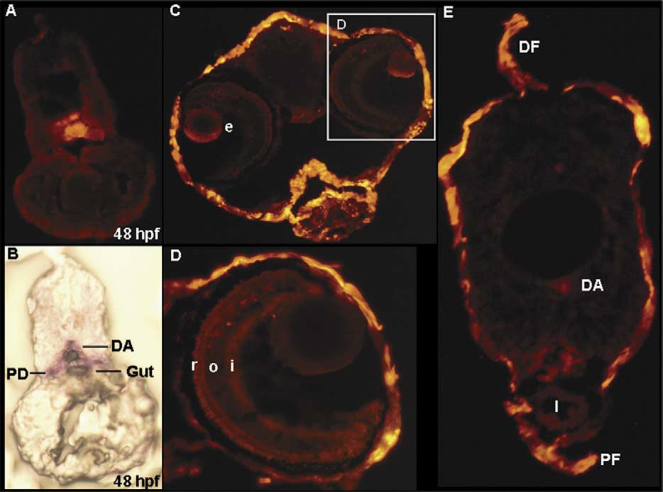
Fig. 3 Cryosections of 48 h postfertilization (hpf) and 7 days postfertilization (dpf) larva from wild-type embryos and Tg(k18(2.9):RFP) transgenic line embryos. (A) Cryosection of the wild-type 48 hpf embryos, revealing that the endogenous k18 was expressed in the dorsal aorta, pronephric duct, and gut. (B) The comparable section from a 48 hpf k18(2.9):RFP transgenic embryo. (C) Head region. (D) Enlarged view of the same section in C. (D) Trunk section. DA, dorsal aorta; DF, dorsal fin; I, intestine; PD, pronephric duct; PF, pelvic fin; e, eye; i, inner plexiform layer; o, outer plexiform layer; r, rods and cones.
Reprinted from Gene expression patterns : GEP, 6(4), Wang, Y.H., Chen, Y.H., Lin, Y.J., and Tsai, H.J., Spatiotemporal expression of zebrafish keratin 18 during early embryogenesis and the establishment of a keratin 18:RFP transgenic line, 335-339, Copyright (2006) with permission from Elsevier. Full text @ Gene Expr. Patterns