
|
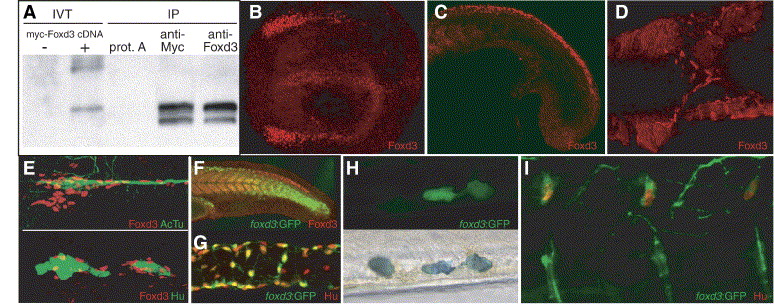
Fig. 1 Expression of Foxd3 protein during zebrafish embryogenesis. (A) Western blot of immunoprecipitated Foxd3 proteins. Left lanes show biotinylated in vitro translation (IVT) reactions with or without cDNA. Right lanes show immunoprecipitation (IP) of in vitro translation reactions with anti-Myc or anti-Foxd3 antibody compared to control where antibody was omitted (prot. A). Biotinylated protein was detected using avidin–HRP. (B–E) Confocal images of Foxd3 antibody staining. (B) Expression in neural crest and floor plate of a 6-somite embryo. Dorsal view, anterior left. (C) Expression in caudal neural crest and somites of an 18 hpf embryo. (D) Expression at 36 hpf in migrating vagal neural crest cells, ventral view with yolk removed. (E) Expression in presumptive glial cells along the posterior lateral line nerve and ganglia. Top panel: red—Foxd3, green—acetylated tubulin. Bottom panel: red—Foxd3, green—Hu. (F) Comparison of Foxd3 immunoreactivity with expression of GFP in the foxd3:GFP transgenic line at 24 hpf. GFP persists in rostral neural crest cells where Foxd3 protein is no longer detectable. (G) Expression of GFP (detected with α-GFP antibody) in a subset of enteric neurons in the dissected intestine of a 96 hpf foxd3:GFP larva. Green—GFP, red—Hu. (H) Expression of GFP in a subset of tail iridophores of a live 72 hpf foxd3:GFP larva. Note GFP-negative iridophore to the left of two GFP-expressing iridophores (iridophore pigments appear dark in this transmitted light image, bottom panel). (I) Confocal image showing expression of GFP (detected with α-GFP antibody) in dorsal root ganglia neurons of a 96 hpf foxd3:GFP larva. Green—GFP, red—Hu.
Reprinted from Developmental Biology, 290(1), Lister, J.A., Cooper, C., Nguyen, K., Modrell, M., Grant, K., and Raible, D.W., Zebrafish Foxd3 is required for development of a subset of neural crest derivatives, 92-104, Copyright (2006) with permission from Elsevier. Full text @ Dev. Biol.