
|
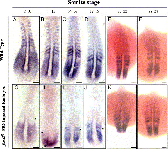
Fig. 6 Whole-mount in situ hybridizations showing gene expression in wild-type (A–F) and the foxd3-MO1-injected embryos (G–L) at different somite stages. In embryos with 8 to 19 somites, myf5 expression in the somites and adaxial cells of foxd3-MO1-injected (10 ng) embryos was much lower than in wild-type embryos. Weak myf5 signals appeared in somites 0 and -1 (arrowheads) and in presomitic mesoderm. Ectopic expression of myf5 was observed in the tail bud (arrow in H). By the 20- to 24-somite stage, myf5 expression patterns in foxd3-MO1-injected and wild-type embryos were similar (E, F vs. K, L). Scale bars: 100 μm.
Reprinted from Developmental Biology, 290(2), Lee, H.C., Huang, H.Y., Lin, C.Y., Chen, Y.H., and Tsai, H.J., Foxd3 mediates zebrafish myf5 expression during early somitogenesis, 359-372, Copyright (2006) with permission from Elsevier. Full text @ Dev. Biol.