
|
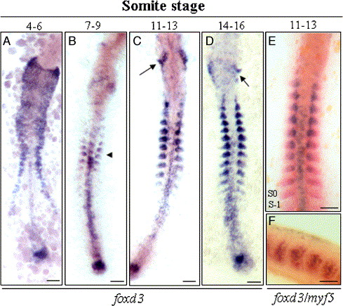
Fig. 4 Temporal and spatial expression of foxd3 in zebrafish embryos at different somite stages. (A) At the 4- to 6-somite stage, foxd3 was transcribed in the floor plate, presumptive neural crest cells, and tail bud. (B) At the 7- to 9-somite stage, foxd3 transcripts were first detected in the somites (arrowhead) and the signals were weak in migrating neural crest cells. In addition to the somitic mesoderm, foxd3 mRNA also was found in the tail bud and posterior premigratory crest. (C, D) At the 11- to 16-somite stage, foxd3 transcription increased incrementally after each pair of somites was formed. foxd3 transcripts in the lateral head were down-regulated, but foxd3 expression was strong in the somites and cranial ganglia posterior to the otic vesicle (arrow). (E, F) In the 11- to 13-somite stage of wild-type embryos, double in situ hybridization using red-labeled myf5 and blue-labeled foxd3 probes was used to show that expressions of these genes were colocalized in the posterior part of the somites. Scale bars: 100 μm.
Reprinted from Developmental Biology, 290(2), Lee, H.C., Huang, H.Y., Lin, C.Y., Chen, Y.H., and Tsai, H.J., Foxd3 mediates zebrafish myf5 expression during early somitogenesis, 359-372, Copyright (2006) with permission from Elsevier. Full text @ Dev. Biol.