
|
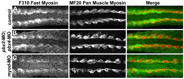
Fig. S4 Fast-muscle differentiation is delayed and reduced in pbx2-MO; pbx4-MO and in myod-MO embryos. (A-C) Antibody staining of fast-muscle myosins with F310 (green in A-C) or pan-muscle myosins with MF20 (red in A-C) in (A) wild-type control, (B) pbx2-MO; pbx4-MO, or (C) myod-MO embryos, all at 16s stage. Somites 4-11 of each embryo are shown in dorsal view, anterior towards the left. MF20 is expressed most strongly in adaxial cells at this stage and appears normal in pbx2-MO; pbx4-MO and in myod-MO embryos. F310 staining is reduced in more posterior somites of pbx2-MO; pbx4-MO and myod-MO embryos.