
Image
|
Figure Caption
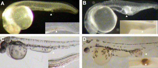
Fig. 1 Morphology of WT (A, 24 hpf; C, 48 hpf) and ChdMO embryos (B, 24 hpf; D, 48 hpf). White arrowhead: Intermediate cell mass. The inserts showed embryos stained with O-dianisidine. Brown color indicates the presence of hemoglobin.
Acknowledgments
This image is the copyrighted work of the attributed author or publisher, and
ZFIN has permission only to display this image to its users.
Additional permissions should be obtained from the applicable author or publisher of the image.
Reprinted from Developmental Biology, 277(1), Leung, A.Y., Mendenhall, E.M., Kwan, T.T., Liang, R., Eckfeldt, C., Chen, E., Hammerschmidt, M., Grindley, S., Ekker, S.C., and Verfaillie, C.M., Characterization of expanded intermediate cell mass in zebrafish chordin morphant embryos, 235-254, Copyright (2005) with permission from Elsevier. Full text @ Dev. Biol.