
|
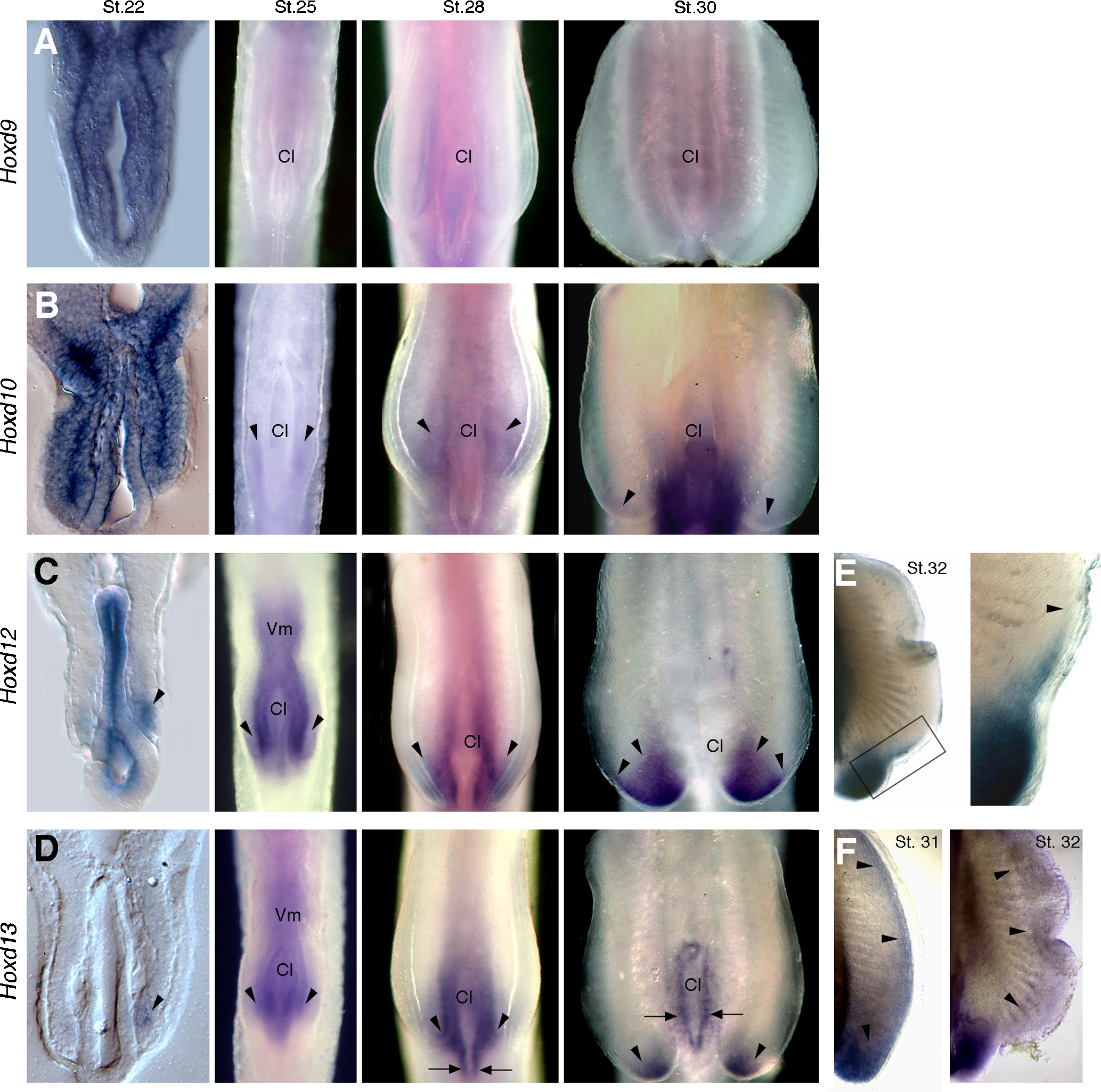
Fig. 3 Expression of Hoxd genes in catshark pelvic fins. Stages of development indicated in the top of each column in A–D and in upper right corner in E and F. Left column shows transverse histological sections at level of cloaca (Cl) and pelvic fins. All other panels show whole mounts in ventral view. (A–D) Whole mount in situ hybridizations showing expression of Hoxd9 (A), Hoxd10 (B), Hoxd12 (C) and Hoxd13 (D). Arrowheads mark expression in pelvic fin buds. Arrows in D mark expression in cloacal epithelium. (E, F) Pelvic fins showing expression of Hoxd12 at stage 32 (E) and Hoxd13 at stages 31 and 32 (F). Boxed area in E is shown in high magnification at right. Arrowheads in E mark anterior limits of expression, and in F they outline the extent of the distal Hoxd13 domain.