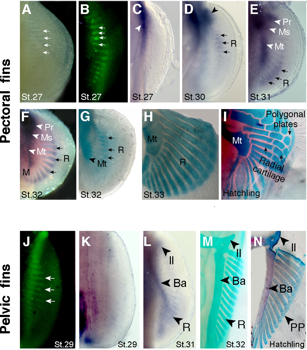
Fig. 1 Endoskeletal development in catshark pectoral and pelvic fins. Ventral views of pectoral (A–I) and pelvic (J–N) fins. Stages (St.) of development indicated at bottom of each panel. (A) Light micrograph of pectoral fin showing gaps in the pectoral fin plate. (B) Acridine orange staining (green fluorescence) shows apoptotic cells in the gaps observed in panel A. Arrows in A and B mark four examples. (C) Sox8 expression marks initiation of chondrogenesis in the pectoral girdle region (arrowhead). Note absence of chondrogenesis in the fin plate at this stage. (D) Sox8 expression marks initiation of chondrogenesis in anterior part of the fin plate, in basal cartilages (arrowhead) and radials (arrows). (E) Sox8 domain prefigures development of the basal cartilages along the anteroposterior axis of the fin: Pr, propterygium; Ms, mesopterygium; Mt, metapterygium; R, radials. Arrows mark expression in the most posterior radials. (F) Sox8 expression in basal cartilages (arrowheads) and in all radials along the anteroposterior axis (subset of radials marked with arrows). (G, H) Alcian green staining of pectoral fins. Note that radials chondrify in domains pre-established by Sox8 expression domains (compare with panels F and G). Chondrified, unsegmented radials are seen in H. (I) Alcian blue and alizarin red stained pectoral fin showing a fully developed cartilaginous endoskeleton at the time of hatching. Note segmentation of proximal radials, intermediate radials and distal polygonal plates (compare panels H and I). (J) Acridine orange-positive cells in gaps of the pelvic fin plate. (K) Sox8 expression marks initiation of chondrogenesis proximal, posterior region of fin. Note absence of chondrogenesis in the fin plate at this stage. (L) Sox8 expression prefigures development of endoskeletal elements in the pelvic fin. Il, iliac process; Ba, basipterygium; R, radials. (M) Alcian green staining of the pelvic fin showing chondrified unsegmented radials. (N) Alcian blue and alizarin red staining of the pelvic fin showing fully developed cartilaginous endoskeleton at hatching. Note segmentation of the radials into distal polygonal plates (PP) and proximal radials (compare panels M and N).
Image
Figure Caption
Acknowledgments
This image is the copyrighted work of the attributed author or publisher, and
ZFIN has permission only to display this image to its users.
Additional permissions should be obtained from the applicable author or publisher of the image.
Full text @ PLoS One

