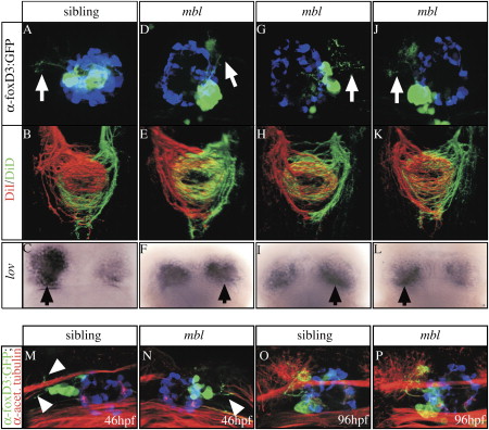
Fig. 5 Epithalamic Asymmetries Are Largely Uncoupled in mbl Embryos (A–P) Dorsal views of (A–L) 4-day-old embryos derived from a mbltm213 x Tg(foxD3:GFP);Tg(lft1:GFP) incross were analyzed for (A, D, G, and J) parapineal migration and projections. The labeling performed and the genotype of the embryos analyzed are indicated on the left and at the top, respectively. (A, D, G, J, M, and N) White arrows mark parapineal projections toward the left or right habenula; pineal cells are pseudocolored in blue. (B, E, H, and K) Axonal projections into the IPN and (C, F, I, and L) lov gene expression (overdeveloped, black arrows mark the side of slightly more intense lov gene expression). The habenulae of all mbl embryos exhibit the projection pattern characteristic for the right habenula, irrrespective of parapineal projections. (M–P) The onset ([M and N]; arrowheads) and targeting (O and P) of parapineal projections is superficially normal in the mbl embryos, irrespective of the migration of parapineal cells. The pineal cells are pseudocolored blue.
Image
Figure Caption
Figure Data
Acknowledgments
This image is the copyrighted work of the attributed author or publisher, and
ZFIN has permission only to display this image to its users.
Additional permissions should be obtained from the applicable author or publisher of the image.
Full text @ Neuron

