
Image
|
Figure Caption
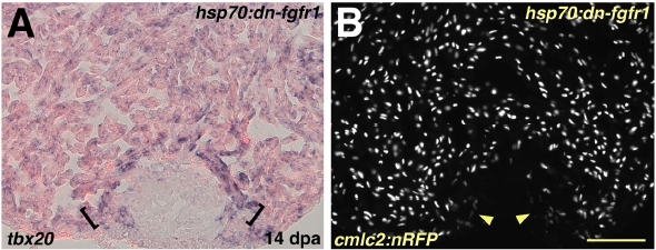
Fig. S5 Myocardial Differentiation per se is Grossly Normal during Fgfr Inhibition (A) Induction of tbx20 in a hsp70:dn-fgfr1 ventricle at 14 dpa. A band of enhanced tbx20 expression at the distal edge of the regenerate adjacent to the wound is indicated by brackets. (B) Ventricular section from a hsp70:dn-fgfr1; cmlc2:nRFP double transgenic animal visualized for RFP fluorescence at 14 dpa. Arrowheads indicate RFPlo CMs. Scale bar = 100 μm.
Figure Data
Acknowledgments
This image is the copyrighted work of the attributed author or publisher, and
ZFIN has permission only to display this image to its users.
Additional permissions should be obtained from the applicable author or publisher of the image.
Reprinted from Cell, 127(3), Lepilina, A., Coon, A.N., Kikuchi, K., Holdway, J.E., Roberts, R.W., Burns, C.G., and Poss, K.D., A Dynamic Epicardial Injury Response Supports Progenitor Cell Activity during Zebrafish Heart Regeneration, 607-619, Copyright (2006) with permission from Elsevier. Full text @ Cell