
|
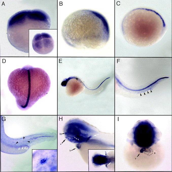
Fig. 2 Expression of zebrafish trpV4 by whole mount in situ hybridization. trpv4 mRNA expression is ubiquitous in blastomeres beginning at the 4-cell stage (A, inset-dorsal view) through to late epiboly. (B) At 75% epiboly, trpv4 shows enhanced expression on the posterior dorsal side of the embryo. At the one somite stage (C) and continuing on until 18 hpf (D), expression is predominately in the notochord with low level expression in the anterior presumptive head region. (E) Head expression becomes enhanced by 24 hpf. (F) At 32 hpf, notochord expression remains strong and trpv4 signal can be detected in the pronephric ducts (black arrow heads). (G) Expression in the notochord is reduced by 40 hpf, decreasing in an anterior to posterior direction. Pronephric duct expression (white arrow heads) persists and staining can be detected in the lateral line organs (black arrow heads, and inset). (H and I) At 3 dpf trpv4 mRNA is highly expressed in brain and head structures. Expression can be seen in the olfactory placodes (op), in chondrogenic tissues including the forming jaw bones (asterisk), pharyngeal arches (white arrow heads), and pectoral fin buds (white arrows, and inset). Expression in the heart (h) is easily detected at this stage, primarily in the ventricle (v) and upper atrium (a).
Reprinted from Gene expression patterns : GEP, 7(4), Mangos, S., Liu, Y., and Drummond, I.A., Dynamic expression of the osmosensory channel trpv4 in multiple developing organs in zebrafish, 480-484, Copyright (2007) with permission from Elsevier. Full text @ Gene Expr. Patterns