
Image
|
Figure Caption
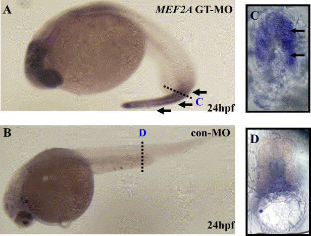
Fig. 3 MEF2A inhibition results in induction of apoptosis in the posterior somites. Embryos were fixed at 24 hpf for TUNEL staining. Black arrows indicate widespread apoptosis in the posterior somites (A, compare to B) (A, n = 20 embryos; B, n = 28 embryos). All embryos are shown in lateral view with the head to the left. Cross-sections of MEF2A GT-MO-injected and control MO-injected embryos are shown in C (n = 5 embryos) and D (n = 5 embryos), respectively. A ubiquitous distribution of apoptosis in the muscles could be found through the cross-sections.
Figure Data
Acknowledgments
This image is the copyrighted work of the attributed author or publisher, and
ZFIN has permission only to display this image to its users.
Additional permissions should be obtained from the applicable author or publisher of the image.
Reprinted from Mechanisms of Development, 123(10), Wang, Y., Qian, L., Dong, Y., Jiang, Q., Gui, Y., Zhong, T.P., and Song, H., Myocyte-specific enhancer factor 2A is essential for zebrafish posterior somite development, 783-791, Copyright (2006) with permission from Elsevier. Full text @ Mech. Dev.