
|
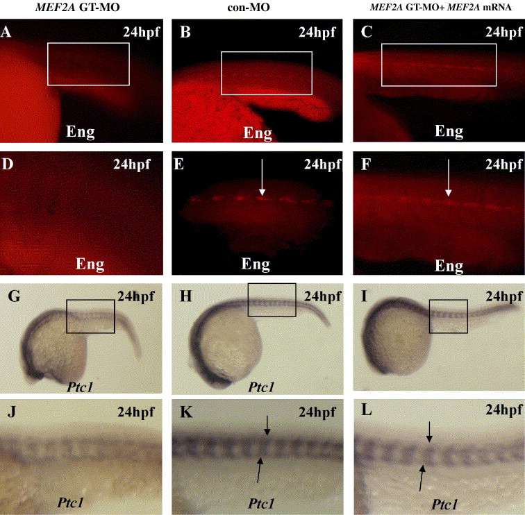
Fig. 2 MEF2A morphants exhibit Hedgehog-associated defects in slow muscle. (A–F) Lateral views of somites 8–13 in whole-mount embryos at 24 hpf. Wild-type embryos show strong Engrailed expression in muscle pioneers (arrow) (B and E) (n = 25 embryos). Engrailed expression in MEF2A morphants is mostly absent, though very weak expression can occasionally be observed (A and D) (n = 20 embryos). At 24 hpf, embryos injected at the 1- to 2-cell stage with 50 pg of MEF2A mRNA plus 10 ng of MEF2A GT-MO (C and F) showed rescue of strong Engrailed expression in the muscle pioneers (arrow) (n = 20 embryos). (G–L) Lateral views of whole-mount embryos with the head to the left at 24 hpf. Wild-type embryos exhibit strong expression of ptc1 (H and K) (n = 30 embryos), while MEF2A morphants (G and J) (n = 20 embryos) show weaker levels of ptc1 expression. Also, embryos injected at the 1- to 2-cell stage with 50 pg of MEF2A mRNA plus 10 ng of MEF2A GT-MO (I and L) showed rescue of strong ptc1 expression (arrow) (n = 20 embryos).
Reprinted from Mechanisms of Development, 123(10), Wang, Y., Qian, L., Dong, Y., Jiang, Q., Gui, Y., Zhong, T.P., and Song, H., Myocyte-specific enhancer factor 2A is essential for zebrafish posterior somite development, 783-791, Copyright (2006) with permission from Elsevier. Full text @ Mech. Dev.