
|
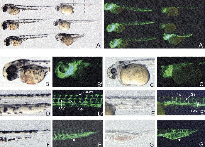
Fig. 6 crim1 morphants display blood vessel defects. (A–G′). Brightfield and darkfield images of Control morpholino (B, B′, D, D′, F, F′) and crim1A-MO (C, C′, E, E′, G, G′) injected into transgenic fli:EGFP embryos and photographed post 48 hpf. In A and A′ panels, the embryos on the left are the Control morpholino injected embryos and on the right are the crim1A-MO injected embryos. Dotted lines in A represent section of embryos used in higher magnification. Note the reduction in GFP intensity in the crim1 morphants. The arrow in D′ shows the presence of the dorsal longitudinal anastomotic vessel (DLAV) and the parachordal vessels (PAV, white arrow) and their absence in E′. These panels (arrows-Se) also highlight the structural differences of the intersegmental vessels (Se) between the Control and crim1 morphants. Arrows in F′ and G′ point to the intermediate cell mass (ICM). Note how this region contains and is surrounded by GFP positive endothelial cells in the crim1 morphants. Scale bar=200 μm.
Reprinted from Mechanisms of Development, 123(4), Kinna, G., Kolle, G., Carter, A., Key, B., Lieschke, G.J., Perkins, A., and Little, M.H., Knockdown of zebrafish crim1 results in a bent tail phenotype with defects in somite and vascular development, 277-287, Copyright (2006) with permission from Elsevier. Full text @ Mech. Dev.