
|
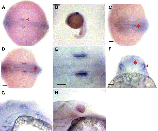
Fig. 4 In situ hybridizations showed cldnj expression in the ear and hindbrain. (A) Seven somite embryo, dorsal view (B) 18 hpf embryo, lateral view low magnification. (C) 18 hpf embryo, dorsal view of head, note weak staining throughout brain marked by red arrow. (D) 18 hpf embryo, dorsal view of otic vesicles. (E) High magnification of 18 hpf embryo, dorsal view of otic vesicles (F) 31 hpf embryo, otic vesicles and hindbrain, posterior view imaging directly through the hindbrain (similar to histological cross-sections in Fig. 2), red arrowhead indicates staining is dorso-lateral in the vesicle. Red arrow marks increased signal of cldnj at the midline of the brain. (G) 31 hpf embryo, lateral view with cldnj staining in the otic placode. (H) 48 hpf embryo, lateral view. Scale bar=100 μm.
Reprinted from Mechanisms of Development, 122(7-8), Hardison, A.L., Lichten, L., Banerjee-Basu, S., Becker, T.S., and Burgess, S.M., The zebrafish gene claudinj is essential for normal ear function and important for the formation of the otoliths, 949-958, Copyright (2005) with permission from Elsevier. Full text @ Mech. Dev.