
|
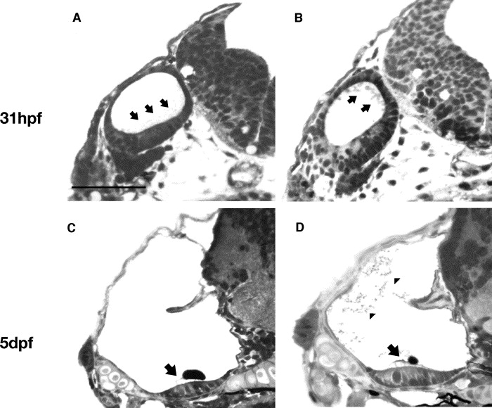
Fig. 2 Histological sections of wildtype and cldnj embryos at 31 hpf and 5 dpf. Orientation is a cross-section of the head with the ectoderm to the left. The otic vesicle is the oval shape at 31 hpf (with the hindbrain to the right) and at 5 dpf it is the large cavity with part of the semicircular canal tissue visible as a projection into the cavity. (A) Histological cross-section through the zebrafish head of wildtype otic vesicle at 31 hpf. Black arrows indicate the ‘haze’ of otolith particles distributed along the ventral-lateral region of the vesicle. (B) cldnj mutant embryo. Otolith material appears as larger granules distributed throughout the vesicle (black arrows). (C) Wildtype ear at 5 dpf. Black arrow indicates the hair cell bundles attached to the fully formed otolith. (D) cldnj mutant ear at 5 dpf. Black arrow points to the normal appearing hair cell bundles attached to a reduced otolith. Arrowheads point to the unincorporated otolith particles still present in the endolymph. Scale bar=100 μm.
Reprinted from Mechanisms of Development, 122(7-8), Hardison, A.L., Lichten, L., Banerjee-Basu, S., Becker, T.S., and Burgess, S.M., The zebrafish gene claudinj is essential for normal ear function and important for the formation of the otoliths, 949-958, Copyright (2005) with permission from Elsevier. Full text @ Mech. Dev.