
|
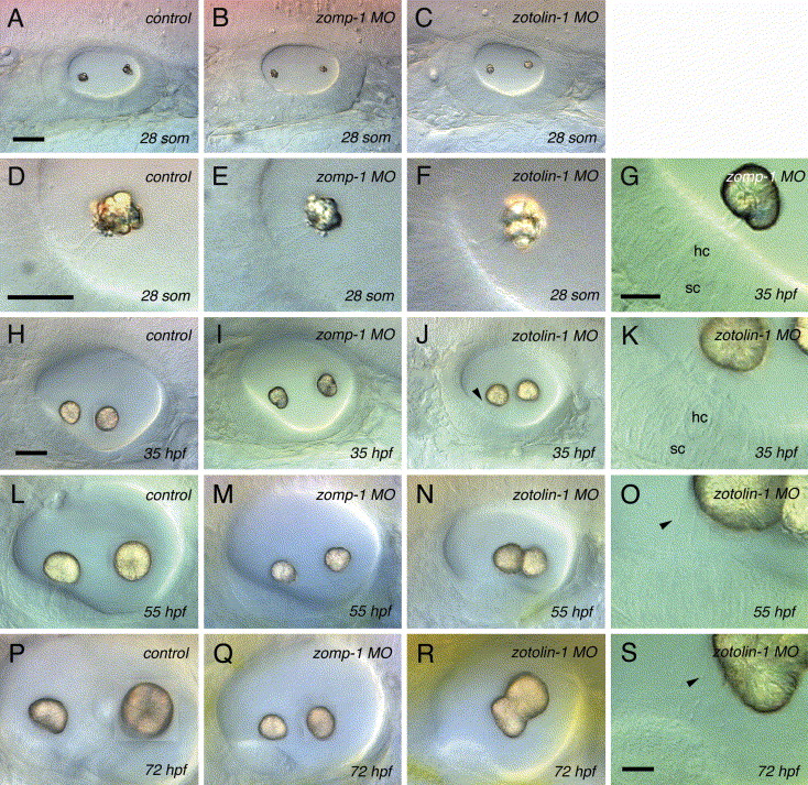
Fig. 3 Otolith phenotypes observed in live control, zomp-1 and zotolin-1 MO-injected embryos by DIC videomicroscopy. Embryos were injected with 4 ng MO. (A–C) 28-somite stage; otolith seeding occurs normally in control MO (A), zomp-1 MO (B) and zotolin-1 MO (C) injected embryos. (D–F) Magnification of the anterior otolith and two tethering kinocilia shown in (A–C). (G–K) At 35 hpf, the otoliths of zomp-1 MO (I) and zotolin-1 MO (J) injected embryos are slightly smaller, and closer to each other, respectively, than those of control MO-injected embryos (H); the anterior otolith of zotolin-1 MO-injected embryos does not stay in contact with the anterior macula (arrowhead in J); (G, K) Anterior macula, otolith and tethering kinocilia of the zomp-1 MO ear shown in (I) and zotolin-1 MO ear shown in (J). (L–N) At 55 hpf, zomp-1 MO and zotolin-1 MO injection cause a ‘small otoliths’ phenotype (M) and a ‘fused otoliths’ phenotype (N), respectively. (O) Magnification of the cilia of the anterior macula shown in (N). At 72 hpf (P–S), the otoliths of control MO (P) and zotolin-1 MO (R) injected embryos keep growing, especially the posterior otolith, while both otoliths of zomp-1 MO-injected embryos stay small (Q). (S) Magnification of the cilia of the anterior macula shown in (R). In panels (A–C), (H), (I), (L), (M), (P) and (Q), the pictures of posterior otoliths are inlayed, from a deeper focal plane, onto the main pictures, which are focused on the anterior otoliths. Through the panels, anterior is to the left, dorsal to the top. Scale bar in (A) indicates 25 μm in (A–C), in (D) indicates 10 μm in (D–F), in (H) indicates 25 μm in (H–J, L–N, P–R), in (G) indicates 10 μm in (G), (K), (O) and (S).
Reprinted from Mechanisms of Development, 122(6), Murayama, E., Herbomel, P., Kawakami, A., Takeda, H., and Nagasawa H., Otolith matrix proteins OMP-1 and Otolin-1 are necessary for normal otolith growth and their correct anchoring onto the sensory maculae, 791-803, Copyright (2005) with permission from Elsevier. Full text @ Mech. Dev.