
Image
|
Figure Caption
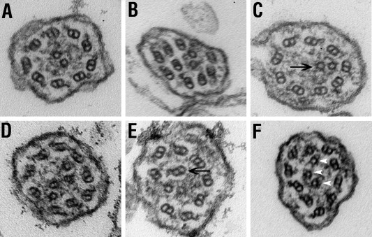
Fig. 7 Ultrastructure of cilia. Images of ultrathin cross-sections through the pronephric cilia of wild-type (A) and lokto237b (B–F) mutant embryos at 5 dpf. (A) Typical 9 + 2 microtubule configuration in wild-type animals. (B–F) Abnormal arrangements of microtubules in lokto237b mutant embryos frequently feature misplaced microtubules (arrows in C and E) or supernumerary microtubule singles (arrowheads in F). Note the irregular shape of membrane in some mutant cilia (B,F).
Figure Data
Acknowledgments
This image is the copyrighted work of the attributed author or publisher, and
ZFIN has permission only to display this image to its users.
Additional permissions should be obtained from the applicable author or publisher of the image.
Reprinted from Mechanisms of Development, 124(7-8), Zhao, C., and Malicki, J., Genetic defects of pronephric cilia in zebrafish, 605-616, Copyright (2007) with permission from Elsevier. Full text @ Mech. Dev.