
Image
|
Figure Caption
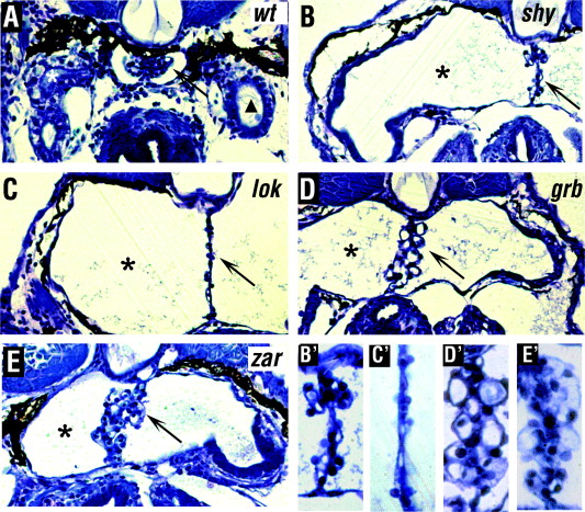
Fig. 2 Histological analysis of the anterior pronephros. Transverse sections through the glomerulus (arrow) and the pronephric tubule (asterisks) in mutant and wild type embryos at 5 dpf. The triangle in (A) indicates the pronephric duct. All mutant strains display a distended pronephric tubule lumen (asterisks in B–E). The glomerulus is stretched out dorso-ventrally in the mutants (arrow in B–E). Enlargements of glomeruli (B′–E′) correspond to mutant strains shown in (B–E). In all panels, dorsal is up.
Figure Data
Acknowledgments
This image is the copyrighted work of the attributed author or publisher, and
ZFIN has permission only to display this image to its users.
Additional permissions should be obtained from the applicable author or publisher of the image.
Reprinted from Mechanisms of Development, 124(7-8), Zhao, C., and Malicki, J., Genetic defects of pronephric cilia in zebrafish, 605-616, Copyright (2007) with permission from Elsevier. Full text @ Mech. Dev.