
|
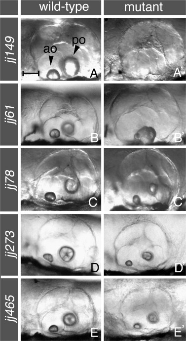
Fig. 2 External phenotypes of otolith mutants. Lateral views of the ear in mutant animals (right column), compared to their wild-type siblings at the same stage (left column). At 144 hpf, no contentjj149 mutant embryos (A′) lack otoliths entirely. At the same stage, the rough edgesjj61 mutation (B′) usually results in one misshapen otolith characterized by a rough appearance, while teeny rocksjj78 (at 144 hpf), pebblesjj273 (at 120 hpf), and condensedjj465 (at 154 hpf) mutants (panels C′, D′, and E′ respectively) feature otoliths reduced in size when compared to their counterparts in wild-type siblings (C–E). Arrowheads in (A) indicate anterior (ao) and posterior (po) otoliths. In all panels, anterior is to the left and dorsal is up. Larvae were photographed using DIC optics. See Table 1 for a further description of phenotypes. Scale bar in (A) applies to all panels, and equals 50 μm.
Reprinted from Mechanisms of Development, 124(7-8), Schibler, A., and Malicki, J., A screen for genetic defects of the zebrafish ear, 592-604, Copyright (2007) with permission from Elsevier. Full text @ Mech. Dev.