
|
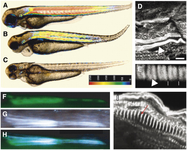
Fig. 3 Expression of steif in the mutant restores myofibrils. (A) uninjected wild-type, (B) steif mutant injected with the BAC clone encoding the steif/unc-45b gene fused with CFP, (C) uninjected steif mutant. The color code on the inserted bar represents the intensity of birefringence covering a five fold range from red (high) to blue (low). (D) F59 staining of BAC-injected steif embryos showing rescue of single fibrils (arrowhead). (E) magnification of panel F. Muscle cells expressing Steif-CFP show normal striation (A-band, arrow head, vertical lines, Z-lines). (F–I) Steif-GFP plasmid injection into steif mutant embryos led to recovery of birefringence and rescue of myofibril organization (F, GFP fluorescence; G, birefringence; H, merge of panels F and G). (I) α-actinin staining showing a rescued fibril in steif mutant embryos (arrow indicates Z-line). Scale bars: 150 μm (A–C); 12 μm (D, F–H); 2 μm (E), 5 μm (I). (A–I) 3-day-old embryos.
Reprinted from Developmental Biology, 308(1), Etard, C., Behra, M., Fischer, N., Hutcheson, D., Geisler, R., and Strähle, U., The UCS factor Steif/Unc-45b interacts with the heat shock protein Hsp90a during myofibrillogenesis, 133-143, Copyright (2007) with permission from Elsevier. Full text @ Dev. Biol.