
Fig. 1
|
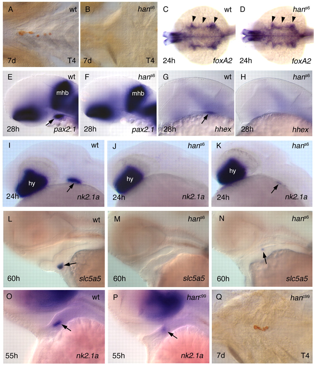
Fig. 1 Thyroid development is impaired in hands off mutant zebrafish embryos. Anterior is to the left. Stages are indicated bottom left, genotype top right and staining/marker bottom right. Arrows show thyroid primordium; arrowheads show pharyngeal endoderm. Ventral (A,B,Q), dorsal (C,D) and lateral (E-P) views are shown. T4 (thyroid hormone) immunostaining (A,B,Q) and in situ hybridisation (C-P). (A,B) In hans6 embryos, no T4-producing follicles are detectable. (C,D) Pharyngeal endoderm appears to be normal in hans6 mutants. (E-K) Expression of thyroid developmental markers. (L-N) The thyroid differentiation marker slc5a5 is expressed in approximately 10% of hans6 mutant embryos. (O-Q) In hanc99 mutants, both primordium and differentiated thyroid are reduced. hy, hypothalamus; mhb midbrain-hindbrain boundary.