
Image
|
Figure Caption
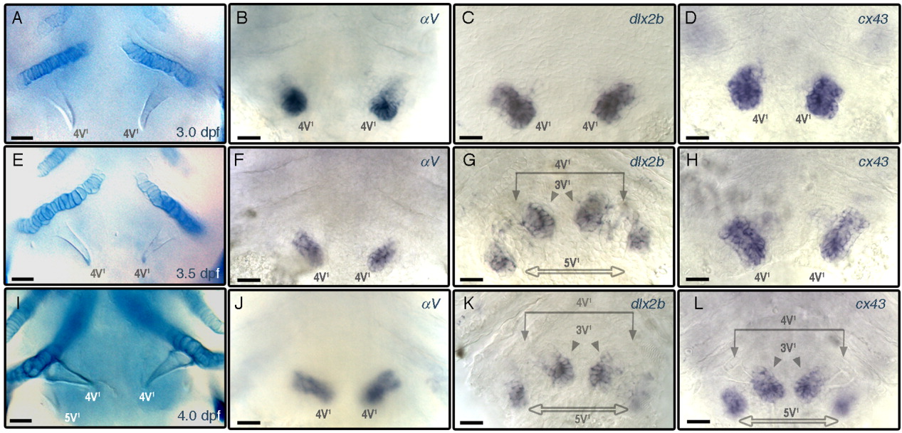
Fig. 6 Spatial and temporal expression patterns of αV, dlx2b, and cx43. A-L: Ventral views of specimens centered at the fifth ceratobranchial, focused at the level of teeth, anterior to the top and oriented as in Figure 5B. A, E, I: Alcian blue stained cartilages. Whole mount ISH specimens probed with αV (B, F, J), dlx2b (C, G, K), and cx43 (D, H, L). Larval stages are identical for each row: 3.0 dpf (A-D); 3.5 dpf (E-H); 4.0 dpf (I-L). Scale bars = 20 μm.
Figure Data
Acknowledgments
This image is the copyrighted work of the attributed author or publisher, and
ZFIN has permission only to display this image to its users.
Additional permissions should be obtained from the applicable author or publisher of the image.
Full text @ Dev. Dyn.