
Fig. 5
|
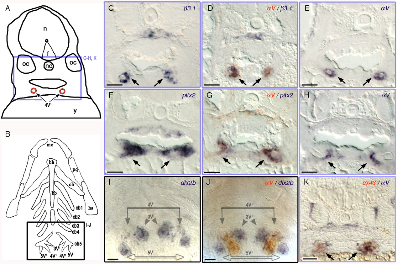
Fig. 5 A-K:αV and β3.1 are expressed in the dental epithelium of the pharyngeal tooth. A: Camera lucida drawing of 3.5 dpf larval transverse sections. Purple box represents the location of the images in the transverse sections in C-H, and K. 4V1, first forming tooth; f, floor plate; n, neural tube; nc, notochord; oc, otic capsule; y, yolk. B: Diagram of the pharyngeal cartilages in 3.5 dpf young larva, ventral view. Black box representing the location of the images in I and J. 3V1/5V1, second forming teeth pair; bb, basibranchials; bh, basihyal; cb, ceratobranchials; ch, ceratohyal; hs, hyosymplectic; mc, Meckel's catilage; pq, platoquadrate. Expression of β3.1 (C,D), αV (D,E, G,H, J,K), pitx2 (F,G), dlx2b (I,J), cx43 (K) in wild type zebrafish. Double ISH samples: (D) αV (orange-red), β3.1 (dark purple); (G) αV (orange-red), pitx2 (dark purple); (J) αV (orange-red), and dlx2b (dark purple); (K) αV (dark purple), and cx43 (orange-red). Scale bars = 20 μm.