
|
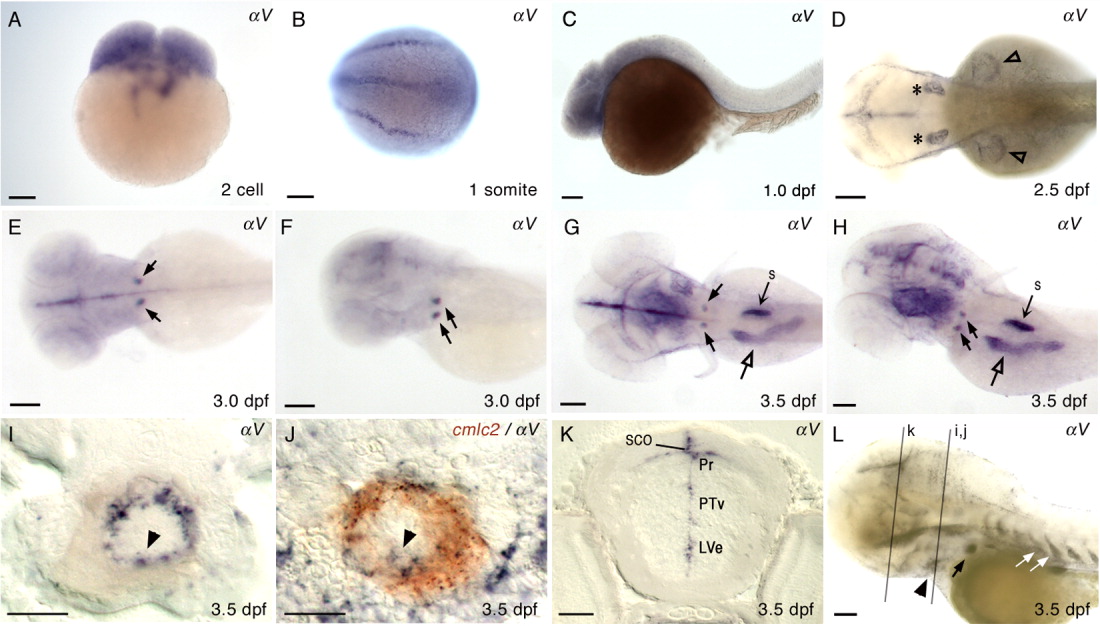
Fig. 3 Expression of αV in wild type zebrafish. Embryonic and larval stages: (A) 2-cell; (B) 1 somite; (C) 1 dpf; (D) 2.5 dpf; (E,F) 3.0 dpf; (G-L) 3.5 dpf. Marked expression fields: ears (asterisk), AER of pectoral fins (open arrowhead), bilateral expression in the deep posterior, ventral pharyngeal region (black arrows), intestine (open arrow), and swim bladder (s). Transverse sections of a 3.5-dpf larva at the level of heart (I, J), and at the level of eye (K). I: αV expression in the ventricle of embryonic heart tube (black arrowhead). J: Double ISH larva, cmlc2 (red), αV (dark purple). K: αV expression in brain: LVe, lateral recess ventricle of hypothalamus; Pr, pretectum; PTv, ventral part of posterior tuberculum; SCO, subcommissural organ. L: Longer staining of 3.5 dpf larva: Intersegmental vessels (white arrow); heart ventricle (black arrowhead). Relative locations for sections in I, J, and K are indicated with gray lines in L. Lateral views of embryos, anterior to the left, are shown in C, F, H, and L. Larva in F and H were slightly tilted. Dorsal views of the embryos, anterior to the left, are shown in B, D, E, and G. Scale bars = 100 μm in A-H and L, and 20 μm in I-K.