
|
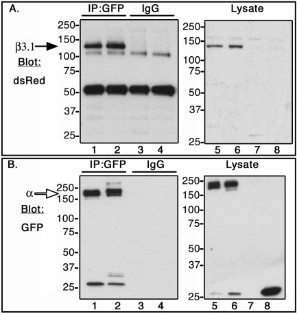
Fig. 2 Co-immunoprecipitation of zebrafish αV, αIIb, and β3.1 subunit in CHO cells. C-terminal α subunits fused to GFP (open arrow, ∼180 kDa), or β3.1 subunit fused to DsRed monomer (black arrow, ∼130 kDa) were expressed in CHO cells. Lysates were incubated with anti-GFP antibody (IP:GFP, lanes 1 and 2), and with isotype control antibody, rabbit IgG (IgG, lanes 3 and 4). Co-immunoprecipitated samples (lanes 1-4), and whole cell lysates (lanes 5-8) were Western blotted with dsRed antibody (A) or GFP antibody (B). CHO cells were transfected in the following order: Lanes 1, 3, and 6, αIIb-GFP + β3.1-dsRed; Lanes 2, 4, and 5, αV-GFP + β3.1-dsRed; Lane 7, DsRed monomer; Lane 8, GFP.