
|
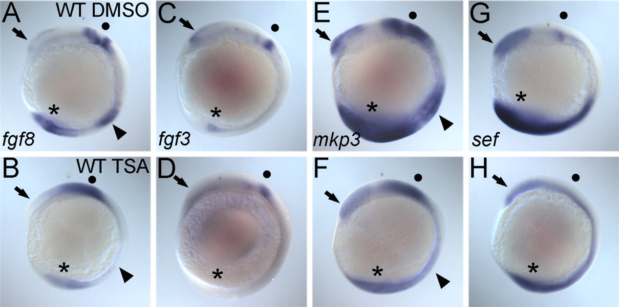
Fig. 9 The proper expression of fgf3 and fgf8 and fgf target genes, sef and mkp3, requires histone deacetylase (HDAC) activity. A-H: All panels show lateral views of whole-mount in situ hybridizations of seven-somite stage WT embryos treated with dimethylsulfoxide (DMSO, A,C,E,G) or 350 nM Trichostatin A (TSA, B,D,F,H) from dome to the seven-somite stage. Probes are shown in the bottom left corner of each wild-type panel. A,B,E,F: The forebrain (arrows) and tail bud (asterisk) expression of all markers is decreased or absent in TSA-treated embryos while the midbrain-hindbrain boundary (MHB) domains (dots) are decreased and diffuse. Both fgf8 (A,B) and mkp3 (E,F) expression in the developing somites (arrowheads) is also lost in TSA-treated embryos.