
|
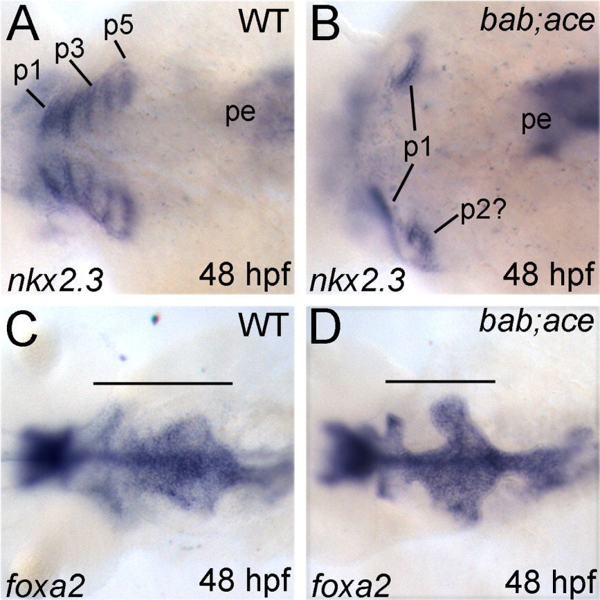
Fig. 7 rerea and fgf8 interact during formation of the endodermal pouches. A-D: All panels show dorsal views, with anterior to the left, of whole-mount in situ hybridizations of wild-type (A,C) and bab;ace (B,D) embryos. Probes are shown in the lower left corner, whereas stages are in the lower right. Combined disruption of rerea and fgf8 leads to loss of the posterior endodermal pouches as seen by nkx2.3 expression (A,B) while medial endoderm (foxa2) remains (C,D). Horizontal bars (C,D) mark the anterior-posterior extent of the pharyngeal endoderm. p1-5, endodermal pouches 1-5; pe, posterior endoderm.